Tethered Spinal Cord
-
Schedule an appointment with the Neurosciences Center
If this is a medical emergency, call 911.
- If you would like an appointment, ask your child’s primary care provider for a referral.
- If you have a referral, call 206-987-2016 or 844-935-3467 (toll-free).
- How to schedule.
If you have any questions, please contact us at 206-987-2016 or 844-935-3467 (toll-free).
-
Find a doctor
- Meet the Neurosciences team.
-
Locations
- Seattle Children’s hospital campus: 206-987-2016 or 844-935-3467 (toll-free)
- Bellevue Clinic and Surgery Center: 206-987-2016 or 844-935-3467 (toll-free)
- Everett: 425-783-6200
- Federal Way: 206-987-2016 or 844-935-3467 (toll-free)
- Olympia: 206-987-2016 or 844-935-3467 (toll-free)
- Tri-Cities: 206-987-2016 or 844-935-3467 (toll-free)
- Wenatchee: 206-987-2016 or 844-935-3467 (toll-free)
-
Refer a patient
- Urgent consultations (providers only): call 206-987-7777 or 877-985-4637 (toll free).
- If you are a provider, fax a New Appointment Request Form (NARF) (PDF) (DOC) to 206-985-3121 or 866-985-3121 (toll-free).
- Send the NARF, chart notes and any relevant documentation to 206-985-3121 or 866-985-3121 (toll-free).
What is a tethered spinal cord?
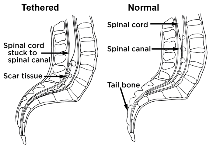
A tethered spinal cord is a spinal cord that is pulled down and stuck, or fixed, to the spinal canal. The spinal cord normally floats free inside the spinal canal.
As a child grows, the spinal cord must be able to move freely inside the spinal canal. If the spinal cord is stuck, it will stretch like a rubber band as a child grows. This can cause lasting damage to the spinal nerves.

-
What causes tethered spinal cord?
Children have tethered spinal cords for many reasons. Tethered spinal cord most often happens in children who have birth defects called myelomeningoceles or lipomyelomeningoceles. Over time, the spinal cords of children with these conditions may become stuck, or tethered, to the myelomeningocele or lipomyelomeningocele. This pulls on the spinal cord as the child grows, causing symptoms.
A child also may have a tethered spinal cord for one of these reasons:
- The very end of their spinal cord is held down more tightly than normal (filum terminale).
- There is a small tract that is not normal, going from the skin into the spinal canal (dermal sinus tract).
- The spinal cord is split into 2 cords near the end (diastematomyelia or diplomyelia).
Tethered Spinal Cord at Seattle Children’s
 Our neurosurgeons have a great deal of experience treating tethered spinal cords and the conditions that can cause them. We know the problem can cause lasting damage to a child’s spinal cord and cause loss of function, like the ability to walk or control their bladder, if the problem is not treated effectively. We do surgery on about 1 child a week who has a tethered spinal cord.
We use neuromonitoring during surgery. This lets our neurosurgeons monitor the nerves and muscles in the lower part of your child’s body during surgery. Neuromonitoring helps neurosurgeons avoid the risk of further damage to your child’s nerves.
Symptoms of Tethered Spinal Cord
Children may have several symptoms of tethered spinal cord, including:
- Back pain or shooting pain in the legs
- Weakness, numbness or problems with muscle function in the legs
- Tremors or spasms in the leg muscles
- Changes in the way the feet look, like higher arches or curled toes
- Loss of bladder or bowel control that gets worse
- Scoliosis or abnormal curve of the spine that changes or gets worse
- Repeated bladder infections
- In a child with an unknown tethered cord, signs on the back such as a fatty mass, dimple, birthmark, tuft of hair or anorectal malformations
Children may also have myelomeningocele symptoms or lipomyelomeningocele symptoms if they have one of these birth defects.
If a tethered spinal cord is not repaired, it can cause lasting nerve damage and loss of function over time.
Diagnosing Tethered Spinal Cord
To diagnose tethered spinal cord, the doctor examines your child, looking for signs and symptoms. Your child most likely will have an MRI (magnetic resonance imaging). This test will help the doctor see inside your child’s body and assess their condition.
The place where the spinal cord ends can differ from child to child. Differences in the place where the cord ends are normal and happen even in children with no spinal cord problems. Also, some children with a tethered spinal cord have only a small tether at the end of the cord.
Often for children with mild signs or symptoms we bring together Neurology, Neurosurgery, Urology and Neurodevelopmentalexperts to evaluate a child and tell whether surgery is needed.
Treating tethered spinal cord
Treatment for a tethered spinal cord usually is surgery to free the spinal cord.
-
Neuromonitoring
Our neurosurgeons use advanced neuromonitoring during surgery. This lets them keep watch on the nerves and muscles of the lower part of your child’s body. It helps neurosurgeons avoid the risk of further damage to your child’s nerves.
-
Laminectomy
To free the spinal cord, neurosurgeons do a surgery called a laminectomy. They remove one or more parts of bones in the spine (vertebrae). This lets them reach the spinal cord, the spinal nerve roots and the thecal sac around the spinal cord.
The neurosurgeons then free the spinal cord by gently cutting, or teasing, it away from the scar tissue or fat. Neurosurgeons use a microscope to help them see the area during the surgery.
After the spinal cord is free, neurosurgeons sometimes apply a patch to the covering of the spinal cord (dura mater). This limits the chances cerebrospinal fluid (CSF) will leak.
-
Future surgeries
Once a child has had surgery to repair the spinal cord or has had the spinal cord freed up (detethered), there is a chance the cord will attach again as the child grows. Some children need more surgeries; this is more common in children who have myelomeningoceles or lipomyelomeningoceles.
Contact Us
If you would like an appointment, ask your child’s primary care provider for a referral. If you have a referral, call 206-987-2016 to make an appointment.
Providers, see how to refer a patient.
If you have questions, contact us at 206-987-2016 or 844-935-3467 (toll free).
-
Schedule an appointment with the Neurosciences Center
If this is a medical emergency, call 911.
- If you would like an appointment, ask your child’s primary care provider for a referral.
- If you have a referral, call 206-987-2016 or 844-935-3467 (toll-free).
- How to schedule.
If you have any questions, please contact us at 206-987-2016 or 844-935-3467 (toll-free).
-
Find a doctor
- Meet the Neurosciences team.
-
Locations
- Seattle Children’s hospital campus: 206-987-2016 or 844-935-3467 (toll-free)
- Bellevue Clinic and Surgery Center: 206-987-2016 or 844-935-3467 (toll-free)
- Everett: 425-783-6200
- Federal Way: 206-987-2016 or 844-935-3467 (toll-free)
- Olympia: 206-987-2016 or 844-935-3467 (toll-free)
- Tri-Cities: 206-987-2016 or 844-935-3467 (toll-free)
- Wenatchee: 206-987-2016 or 844-935-3467 (toll-free)
-
Refer a patient
- Urgent consultations (providers only): call 206-987-7777 or 877-985-4637 (toll free).
- If you are a provider, fax a New Appointment Request Form (NARF) (PDF) (DOC) to 206-985-3121 or 866-985-3121 (toll-free).
- Send the NARF, chart notes and any relevant documentation to 206-985-3121 or 866-985-3121 (toll-free).