Current Projects
Uncovering a gene’s role in the development of the trachea
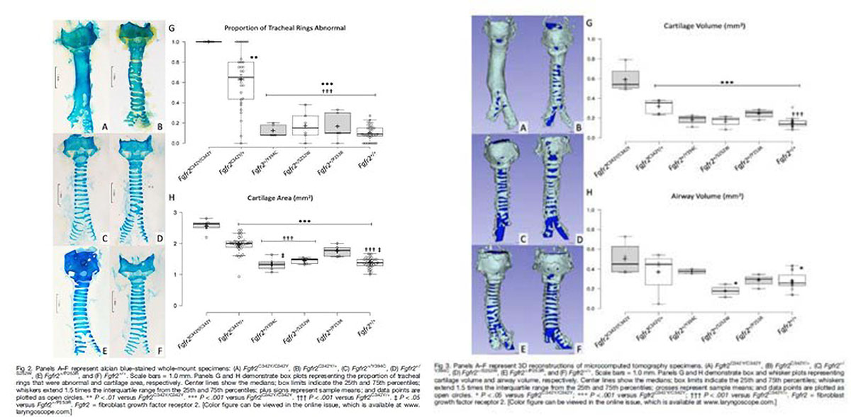
Mutations in fibroblast growth factor receptor 2 gene (FGFR2) cause tracheal cartilaginous sleeve, a congenital malformation associated with several rare craniofacial conditions, such as syndromic craniosynostosis. Patients born with this malformation don’t have typical tracheal cartilaginous rings. Instead, their trachea is an uninterrupted segment of cartilage. Without these cartilaginous rings, the airway does not have a normal function and the patients struggle to breathe.
Through a research collaboration with the Millen Lab, we’re developing a mouse model to better understand tracheobronchial development, the relationship of FGFR2 with tracheal cartilaginous sleeve phenotypes and the factors impacting patients with tracheal malformations. Eventually, we’re aiming to use this model to develop novel diagnostic modalities and personalized treatment approaches for these patients.
Developing a new way to better diagnose and quantify upper airway obstructions
Surgical treatments for upper airway obstruction are often very invasive procedures that carry a high risk of complications, and they’re not always successful in alleviating airway obstruction. This can lead to multiple surgeries and high morbidity for these infants and children.
Today, candidates are selected for these procedures based on several qualitative and subjective diagnostic modalities. Our lab is collaborating with the Multiphase & Cardiovascular Flow Lab at the University of Washington to develop a novel quantitative diagnostic modality that may provide a more accurate and methodical assessment of upper airway obstruction.
 We are integrating dynamic CT scan with computational fluid dynamic analysis to assess upper airway obstruction using quantitative metrics. With this data, we’re working to identify the sites of obstruction, quantify the severity of the obstruction on airway flow and risk stratify patients based on their mild, moderate or severe obstruction.
Our goal is to develop a protocol to better diagnose patients and objectively determine the patients that are the best candidates for surgical treatment and which surgical treatment would be most beneficial.