Diagnostics and Therapeutics for Sepsis
Understanding how mast cell proteases influence mediators generated during the septic response
Technology Overview
Sepsis is a complex and potentially life-threatening condition that occurs when a person’s inflammatory response to infection is dysregulated. There are more than 1 million cases of sepsis per year in the United States, mostly in the elderly, children, and in individuals who are immunocompromised, with mortality rates reaching as high as 50%. Because no specific biomarker for sepsis has been identified, diagnosis is often delayed; thus there is urgent need for biomarkers that promote or restrain the inflammatory response that occurs in sepsis.

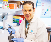
Dr. Adrian Piliponsky
The Piliponsky Lab is investigating the importance and functions of mast cells in inflammatory disorders including sepsis, and testing whether these cells can be manipulated to prevent or control these processes. Mast cells are immune cells widely distributed in the body’s tissues that play a role in allergies and anaphylaxis. They are increasingly recognized as key regulatory cells involved in inflammation, including sepsis, with both pro-inflammatory and anti-inflammatory roles, depending on the pathological context and the mast cell phenotype.
Dr. Adrian Piliponsky uses a combination of in vitro and in vivo approaches to investigate specific mast cell functions and products in diverse models of inflammation, including mouse models that genetically lack mast cells and can undergo engraftment with wild-type or genetically altered mast cells. His work with various animal models confirms that mast cells can significantly influence multiple features of acute and chronic inflammatory responses, through diverse effects that can either promote or suppress aspects of these responses. Some models have correlated the presence and activation of mast cells with exacerbated inflammation and disease progression, whereas others indicate mast cells play a protective role in some contexts such as in wound healing and in some cardiovascular disease and cancers.
The Piliponsky Lab is currently focused on understanding how mast cell proteases can shape the profile of mediators generated during the septic response. These studies will lay the groundwork for future projects aimed at exploring the therapeutic potential of newly-identified pathways and mediators and therefore may prevent the dysregulated inflammatory response that is associated with multiple organ dysfunction and death in severe sepsis.
Stage of Development
- Pre-clinical in vivo
- Pre-clinical in vitro
- Pre-clinical ex vivo
Partnering Opportunities
- Collaborative research opportunity
- Sponsored research agreement
- Consultation agreement
Publications
- Piliponsky AM, Lahiri A, Truong P, Clauson M, Shubin NJ, Han H, Ziegler SF. Thymic stromal lymphopoietin improves survival and reduces inflammation in sepsis. Am J Respir Cell Mol Biol. First published online 2 Mar 2016 as DOI: 10.1165/rcmb.2015-0380OC
- Gendrin C, Vornhagen J, Ngo L, Whidbey C, Boldenow E, Santana-Ufret V, Clauson M, Burnside, K, Galloway DP, Waldorf KA, Piliponsky AM, Rajagopal L. Mast cell degranulation by a hemolytic lipid toxin decreases GBS colonization and infection. Science Advances. 2015: E1400225
- Schäfer B, Piliponsky AM, Oka T, Song CH, Gerard NP, Gerard C, Tsai M, Kalesnikoff J, Galli SJ. Mast cell anaphylatoxin receptor expression can enhance IgE-dependent skin inflammation in mice. J Allergy Clin Immunol. 2013; 131(2):541-548.e1-9.
- Piliponsky AM, Chen CC, Rios EJ, Treuting PM, Lahiri A, Abrink M, Pejler G, Tsai M, Galli SJ. The chymase mouse mast cell protease 4 degrades TNF, limits inflammation, and promotes survival in a model of sepsis. Amer J Pathol. 2012; 181(3):875-886.
- Shubin NJ, Glukhova VA, Clauson M, Truong P, Abrink M, Pejler G, White NJ, Deutsch GH, Reeves SR, Vaisar T, James RG, Piliponsky AM. Proteome analysis of mast cell releasates reveals a role for chymase in the regulation of coagulation factor XIIIA levels via proteolytic degradation. J Allergy Clin Immunol 2017;139(1):323–334.
Learn More
To learn more about partnering with Seattle Children’s Research Institute on this or other projects, email the Office of Science-Industry Partnerships.